
[ad_1]
Case report
An 81-year-old woman initially presented to a dermatology clinic with recurrent petechiae, bruising, sometimes hemorrhagic blisters that did not rupture easily, all over her body for 6 years with no pain or itching. Almost at the same time, the patient found an abnormality of the nails, erythematous and swollen palms, papules on the buttocks and a gradual loosening of the skin. She has had no recent significant weight change.
Physical examination showed multiple petechiae, ecchymoses with negative Nissl’s sign all over the body, abnormal skin wrinkles and laxity, symmetrical periorbital edema accompanied by purpura, and multiple waxy, smooth, brown-colored papules on the face and the anterior cervical region (Pictures 1 to 4), hard warty plaque and papules on the left buttock (Figure 5), multiple nail dystrophy, palmo-digital erythematous swelling.
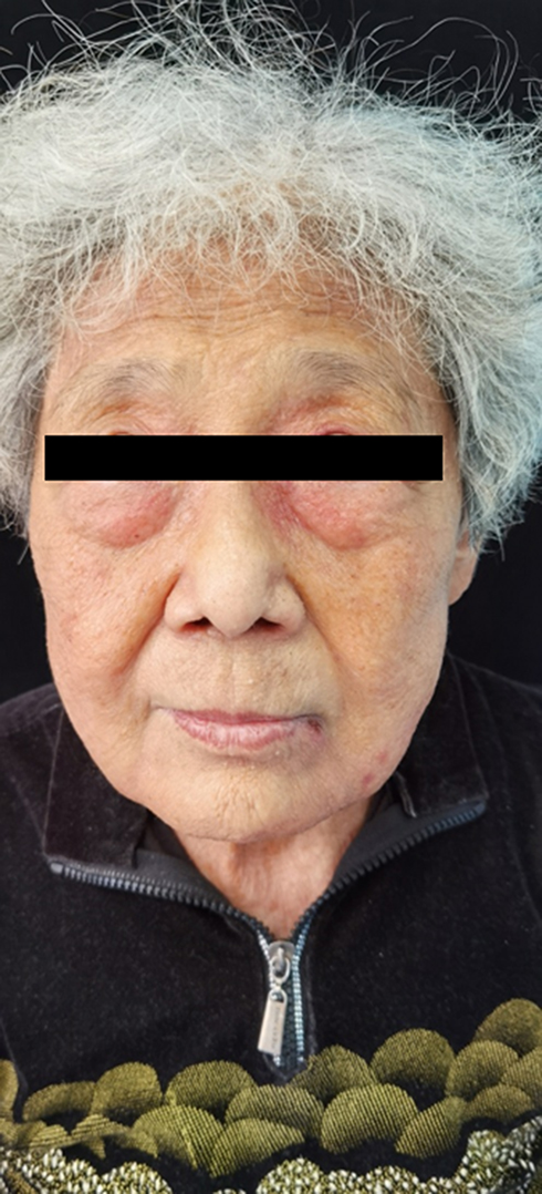 |
Figure 1 A symmetrical periorbital edema accompanied by purpura and multiple smooth, waxy, flesh-colored or brown papules were found on the patient’s face. |
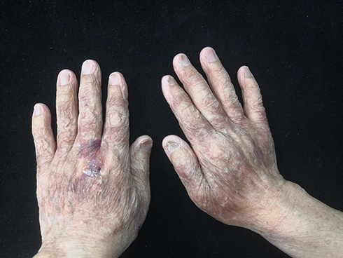 |
Figure 2 Purpura, petechiae and ecchymosis of the skin and dystrophy of the nails. |
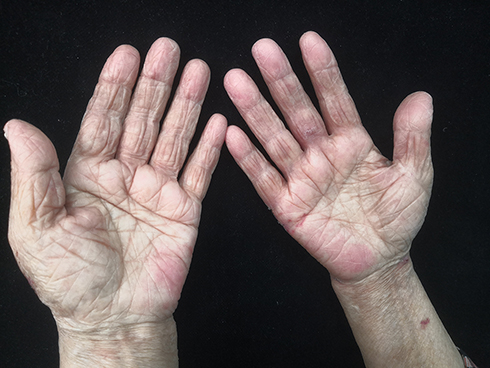 |
picture 3 Palmo-digital erythematous swelling. |
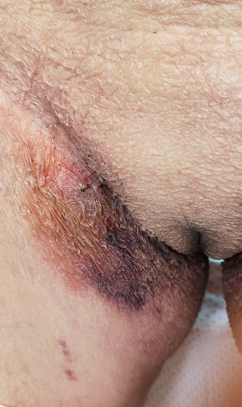 |
Figure 4 The patient’s right groin had a bruise with no apparent cause. |
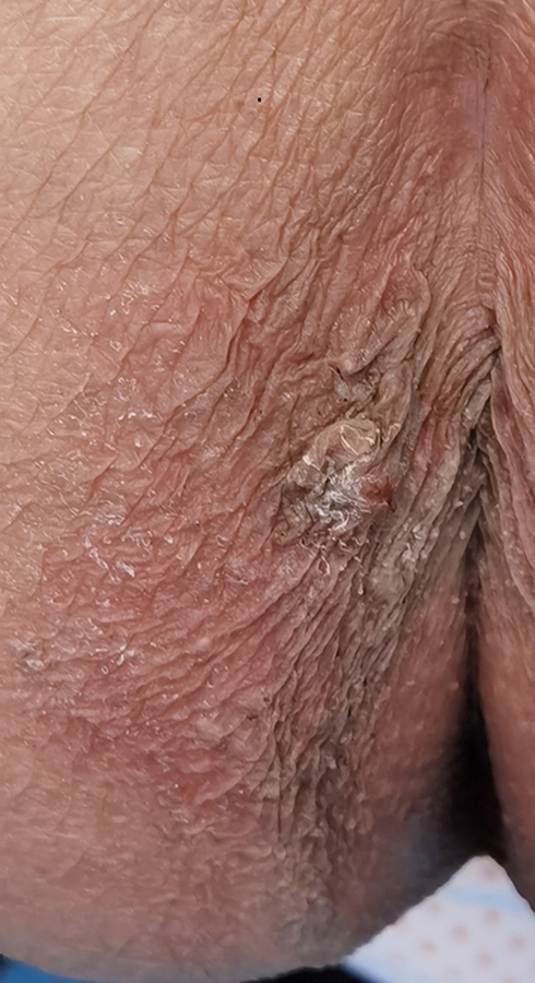 |
Figure 5 There was a hard warty plaque on the left buttock at 3 o’clock. Bleeding may occur after crushing. |
Blood tests showed serum IgG 3.73 g/L (8.60–17.40 g/L), IgA 34.40 g/L (1.00–4.20 g/L), IgM 0, 08 g/L (0.50–2.80 g/L), kappa light chain 3.45 g/L(6.29–13.50g/L), lambda light chain 22.90 g/L(3, 13–7.23g/L), κ/λ 0.15(1.53–3.29), coagulation factor X 48.70% (77–131%).
Protein electrophoresis and immunofixation electrophoresis showed albumin 48.2% (54.0–65.7%), β2-globulin 32.3% (8.2–13.8 %), γ-globulin 1.9% (10.6–23.5%), monoclonal IgA-α and monoclonal λ light chain. The M protein was positive and was of the IgA-λ type. Serum free light chains showed κ 6.2 mg/L (3.30–19.40 mg/L), λ 307.5 mg/L (5.71–26.3 mg/L), Fκ /Fλ 0.020 (0.26–1.65) and dFLC 301.3 mg/L /L. Urine examination showed protein quantification 0.32 g/L, albumin 98.3%, monoclonal IgA-α positive, monoclonal λ, Bence-Jones type λ protein, λ light chain 7.44 mg/dl (0.00-5.00 mg/dl) and 24 hour total urine Quantification of λ light chains 130mg.
The radiological examination showed no bilateral abnormality of the femur and the humerus. The CT scan showed no obvious puncture-like bone destruction in the skull, no obvious bone destruction throughout the spine, ribs, sternum, and pelvic bones, but cervical and lumbar degeneration.
Bone marrow biopsy showed hypercellularity with plasma cell hyperplasia, consistent with multiple myeloma.
Histopathological features of the abdominal skin showed that eosinophilic, morphed, and fissured material was present in the dermis that was positive for Congo red staining (Numbers 6 And 7). Electron microscopy showed that sheets of fine fibers 10 nm in diameter were disorderly distributed in the dermis (Picture 8). The features were consistent with amyloidosis.
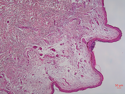 |
Figure 6 Histological characteristics. |
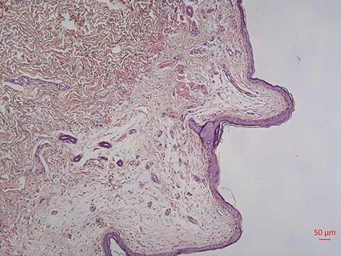 |
Picture 7 Congo red coloring (+). |
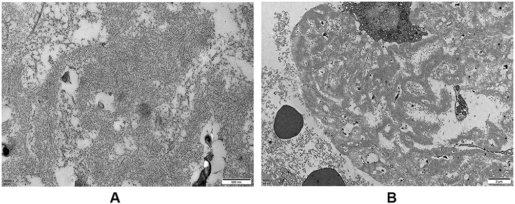 |
Picture 8 (A And B) Electron microscopy showed that sheets of fine fibers with a diameter of about 10 nm were disorderly distributed in the dermis, which is consistent with amyloidosis. |
The patient was diagnosed with multiple myeloma and systemic amyloidosis associated with multiple myeloma. After treatment with the BD regimen (a regimen of bortezomib in combination with dexamethasone), no significant improvement in skin lesions was observed. The patient refused to continue treatment for financial reasons.
Discussion
Immunoglobulin (AL) light chain amyloidosis (known as primary amyloidosis in history) is a rare disease, and the exact incidence is still unknown. In the United States, the incidence appears to be stable at around 9 to 14 cases per million person-years.1–3 The elderly are more susceptible to AL amyloidosis. As with other plasma cell dyscrasias, age-specific incidence rates increase with each decade of life after age 40.1 AL amyloidosis in which the fibrils are composed of fragments of monoclonal light chains. Affected patients may suffer from amyloidosis alone, or may be accompanied by other plasma cell dyscrasias (multiple myeloma, Waldenstrom’s macroglobulinemia). This part of AL amyloidosis is related to multiple myeloma, in which the deposited light chains originate from plasma cells in the bone marrow, resulting in extensive organ deposition and dysfunction.4–6 There are complex interactions between proteins derived from immunoglobulins, in particular the light and heavy chains, and the components of elastic tissues, leading to different types of alteration of the latter.7 Therefore, AL amyloidosis could have various dermatology clinical manifestations, including purpura, waxy thickening, ecchymosis, and cutis laxa.7,8
Amyloid purpura is suspected to be associated with a reduction in coagulation factor X, but this link is not established. It was observed that in 36 patients, low levels of factor X activity were observed in approximately 1 in 4 samples.9 Since AL amyloidosis is a clonal plasma cell disease, it is treated with chemotherapy to eradicate the underlying clone. AL amyloidosis should be differentiated from other forms of amyloidosis (eg, AA amyloidosis, ATTRmt amyloidosis, and ATTRwt amyloidosis) because the latter are nonneoplastic and will not benefit from chemotherapy. Compared to normal conditions, MM cells may express lower or higher levels of microRNAs (miRs), which are used as tumor suppressors or oncogenes. Since the expression of tumor suppressor miRs is low in cancer, it may offer therapeutic benefits to restore their normal levels through a miR replacement strategy.ten
Conclusion
Our patient presented with purpura, smooth, waxy or brown papules, verrucous plaque, hemorrhagic blisters and nail dystrophy in at least five different forms of skin lesions at the same time. We confirmed the presence of amyloid fibrils using Congo red staining for identity. We also observed under an electron microscope, and found sheets of fine fibers with a diameter of about 10 nm distributed in a disordered manner in the dermis, compatible with amyloid fibrils. Multiple myeloma was diagnosed by bone marrow biopsy, according to the updated criteria for the diagnosis of multiple myeloma by the International Myeloma Working Group, so the final diagnosis for this patient was systemic amyloidosis associated with multiple myeloma .
We report a case of systemic amyloidosis associated with multiple myeloma, in which the simultaneous presentation of multiple skin lesions at different sites in a patient is clinically rare. It is difficult to identify the same dermatosis because of the diversity of the morphology of the skin lesions. Regarding skin manifestations, since the morphology of the rash depends on the site of amyloid deposition, if it deposits in small vessels of the skin, the skin lesions appear as petechiae, bruises and purpura; if deposited in the superficial layer of the dermis, it will present with waxy papules; and if deposited in the dermal elastic tissue, it will exhibit cutis laxa. Initial skin manifestations included multiple skin manifestations in addition to typical signs of amyloidosis. Petechiae and bruising were the first symptoms in our patient.
Ethics statements
Written informed consent for publication of their clinical details and clinical images was obtained from the patient. No institutional approval was required. This case report was prepared in accordance with CARE guidelines.11
Disclosure
The authors report no conflict of interest in this work.
The references
1. Kyle RA, Linos A, Beard CM, et al. Incidence and natural history of primary systemic amyloidosis in Olmsted County, Minnesota, from 1950 to 1989. Blood. 1992;79(7):1817. doi:10.1182/sang.V79.7.1817.1817
2. Quock TP, Yan T, Chang E, et al. Epidemiology of AL amyloidosis: a real-world study using US claims data. Adv of blood. 2018;2(10):1046. doi:10.1182/bloodadvances.2018016402
3. Kyle RA, Larson DR, Kurtin PJ, et al. Incidence of AL amyloidosis in Olmsted County, Minnesota, from 1990 to 2015. Mayo ClinProc. 2019;94(3):465–471. doi:10.1016/j.mayocp.2018.08.041
4. Hazenberg BP. Amyloidosis: a clinical overview. Rheum Dis Clin North America. 2013;39(2):323–345. doi:10.1016/j.rdc.2013.02.012
5. Rosenzweig M, Landau H. Light chain (AL) amyloidosis: update on diagnosis and management. J Hematol Oncol. 2011;18(4):47. doi:10.1186/1756-8722-4-47
6. Bahlis NJ, Lazarus HM. AL amyloidosis associated with multiple myeloma: is a particular therapeutic approach justified? Bone marrow transplant. 2006;38(1):7–15. doi:10.1038/sj.bmt.1705395
7. Ruszczak Z, Wozniak L. Localized nodular cutaneous amyloidosis with anetoderma. dermatologist. 1961;12:254–259.
8. Eder L, Bitterman H. Image in clinical medicine. Amyloid purpura. N English J med. 2007;356(23):2406. doi:10.1056/NEJMicm061510
9. Gamba G, Montani N, Anesi E, et al. Coagulation alterations in primary systemic amyloidosis. Haematological. 2000;85(3):289–292.
ten. Desantis V, Saltarella I, Lamanuzzi A, et al. MicroRNA-based nano-strategies as a new therapeutic approach in multiple myeloma to overcome disease progression and drug resistance. Int J Mol Sci. 2020;21(9):3084. doi:10.3390/ijms21093084
11. Riley DS, Barber MS, Kienle GS, et al. CARE Guidelines for Case Reports: Explanation and Elaboration Document. JclinEpi. 2017;89:218–235.
|
Sources 2/ https://www.dovepress.com/a-case-of-multiple-myeloma-associated-systemic-amyloidosis-with-multip-peer-reviewed-fulltext-article-CCID The mention sources can contact us to remove/changing this article |
[ad_2]